Cells & molecules
T-cell activation
Immunology · Immunological synapse
 Open the interactive 3D model →
How does one T cell, out of millions, know it has found the exact germ it was made to fight?
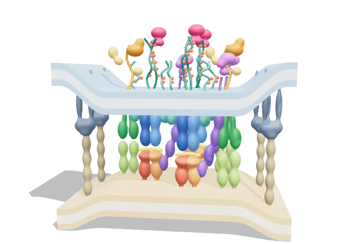
Helper T cells are white blood cells that direct your immune defences. Other cells show them small pieces of germs held in display molecules called MHC. When a T cell’s receptor fits one of those pieces and the presenting cell also gives an “OK” signal, a chain reaction inside the T cell switches it on, and it starts making a messenger called IL-2 and multiplying.
A helper T cell is activated when its T-cell receptor recognises a specific peptide bound to MHC class II on an antigen-presenting cell, while CD4 and the co-stimulatory receptor CD28 add their signals across a specialised contact zone called the immunological synapse. Phosphorylation of CD3 ITAMs starts a kinase cascade through Lck, ZAP-70 and LAT that raises intracellular Ca²⁺ and switches on genes such as IL-2, driving clonal expansion.
Key facts
- TCR–peptide–MHC affinity
- K_d ≈ 1–100 µM (weak), bond lifetime ≈ seconds
- Synapse gap at TCR–pMHC
- ≈ 13–15 nm
- Synapse gap at LFA-1–ICAM-1
- ≈ 40 nm
- ITAMs per TCR–CD3 complex
- 10 (6 on ζζ, 2 on ε, 1 on γ, 1 on δ)
- Antigen sensitivity
- as few as 1–10 peptide–MHC complexes can trigger a response
- Peptide length in MHC II
- ≈ 13–25 residues (open-ended groove)
Parts you can explore (13)
- APC membrane
- MHC class II–peptide
- T-cell membrane
- TCR αβ
- CD3 complex
- CD4 co-receptor
- Lck
- ZAP-70
- CD28–B7 co-stimulation
- LFA-1–ICAM-1 adhesion
- LAT signalosome
- Ca²⁺ signal
- IL-2 cytokine
In the 3D model
Rotate and take apart the model, follow a guided tour of what each part does and why it’s needed, change it with a live slider, and test yourself with a short quiz. Free, no sign-up.
Open T-cell activation in 3D →
OpenArc Models’ models are simplified teaching schematics. Terms · Privacy