Human body
Chemical synapse
Neuroscience · Synaptic transmission
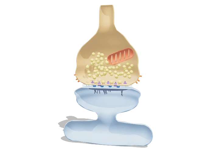 Open the interactive 3D model →
If nerve cells don’t actually touch, how does a message get from one to the next?
Nerve cells pass messages at junctions called synapses, where a tiny gap separates one cell from the next. When an electrical signal reaches the end of the first cell, it releases a puff of chemical messenger into the gap. The messenger drifts across and opens channels in the next cell, starting a new electrical signal there.
At a chemical synapse an action potential opens voltage-gated Ca²⁺ channels in the presynaptic terminal, and the Ca²⁺ influx triggers SNARE-mediated fusion of transmitter-filled vesicles with the membrane. The released transmitter crosses a 20 nm cleft, opens ligand-gated ion channels in the postsynaptic membrane and is then rapidly cleared, so each spike produces a brief, graded postsynaptic potential.
Key facts
- Synaptic cleft width
- ≈ 20 nm (15–25 nm)
- Synaptic vesicle diameter
- ≈ 40 nm; ≈ 2,000–4,000 glutamate molecules each
- Synaptic delay
- ≈ 0.5–1 ms from presynaptic spike to postsynaptic current
- Release probability
- ≈ 0.1–0.9 per vesicle site per spike (often < 0.5 in cortex)
- Synapses in the human brain
- ≈ 10¹⁴–10¹⁵
- Peak cleft glutamate
- ≈ 1 mM, cleared within ≈ 1–2 ms
Parts you can explore (12)
- Presynaptic terminal
- Synaptic vesicles
- SNARE complexes
- Voltage-gated Ca²⁺ channels
- Synaptic cleft
- Neurotransmitter
- Ionotropic receptors
- Postsynaptic density
- Postsynaptic membrane
- Reuptake transporters
- Mitochondrion
- Ca²⁺ and Na⁺ ions
In the 3D model
Rotate and take apart the model, follow a guided tour of what each part does and why it’s needed, change it with a live slider, and test yourself with a short quiz. Free, no sign-up.
OpenArc Models’ models are simplified teaching schematics. Terms · Privacy