Human body
Skeletal muscle
Anatomy · Muscle tissue hierarchy
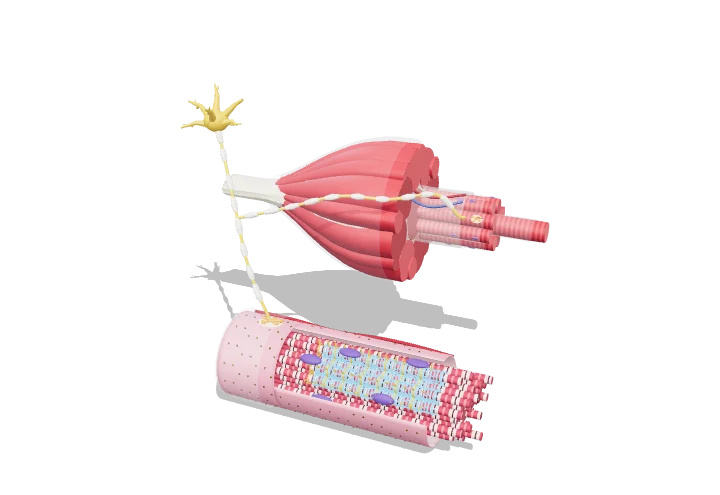 Open the interactive 3D model →
What actually happens inside your arm in the split second between deciding to move and moving?
A muscle is built like a rope: bundles inside bundles inside bundles, down to tiny strands that can slide past each other. A nerve sends an electrical signal that spreads into every muscle cell and releases calcium. The calcium lets the strands grab and pull, so the whole muscle shortens and tugs on the bone.
A skeletal muscle is a nested hierarchy — muscle, fascicle, fibre, myofibril, sarcomere — held together by continuous connective-tissue sheaths that deliver force to the tendon. Each contraction begins with an action potential at the neuromuscular junction that travels into the fibre along T-tubules and releases Ca²⁺ from the sarcoplasmic reticulum, letting the sarcomeres shorten.
Key facts
- Fibre size
- ≈ 10–100 µm wide; millimetres to > 10 cm long
- Myofibril diameter
- ≈ 1–2 µm
- Resting sarcomere length
- ≈ 2.0–2.5 µm (optimal overlap ≈ 2.0–2.2 µm in human muscle)
- T-tubules per sarcomere
- 2 in mammals (at the A–I junctions); 1 at the Z-disc in frogs
- Resting membrane potential
- ≈ −90 mV
- Single twitch duration
- ≈ 25–100 ms (fast vs slow fibres) after a 1–2 ms action potential
Parts you can explore (14)
- Tendon
- Epimysium
- Fascicles
- Perimysium
- Muscle fibres
- Endomysium
- Sarcolemma
- Myofibrils
- Sarcoplasmic reticulum
- T-tubules
- Nuclei
- Motor neuron
- Neuromuscular junction
- Capillaries
In the 3D model
Rotate and take apart the model, follow a guided tour of what each part does and why it’s needed, change it with a live slider, and test yourself with a short quiz. Free, no sign-up.
OpenArc Models’ models are simplified teaching schematics. Terms · Privacy