Cells & molecules
Sarcomere
Physiology · Muscle contraction
 Open the interactive 3D model →
When you lift a cup, what actually moves inside your muscle?
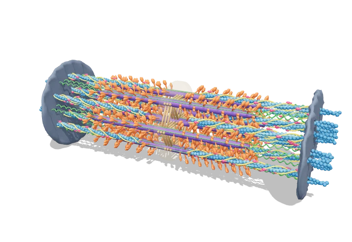
Muscles are built from millions of tiny repeating units called sarcomeres, lined up end to end. Each one holds two kinds of protein strands that overlap like interlaced fingers. When calcium arrives, little “heads” on the thick strands grab the thin strands and pull them inward, so every unit gets a bit shorter — and the whole muscle contracts.
The sarcomere is the repeating contractile unit of striated muscle, bounded by Z-discs and built from overlapping thick (myosin) and thin (actin) filaments arranged in a hexagonal lattice. When Ca²⁺ switches on the thin filaments, myosin cross-bridges cycle on actin and the filaments slide past each other, shortening the sarcomere without either filament changing length.
Key facts
- Resting sarcomere length
- ≈ 2.0–2.5 µm (frog optimum 2.0–2.25 µm)
- Thick filament
- ≈ 1.6 µm long, ~300 myosin II molecules
- Thin filament
- ≈ 1.0 µm (frog; longer in many mammalian muscles)
- Lattice
- 6 thin around each thick; ~2 thin per thick
- Myosin head repeat
- crowns every 14.3 nm; helix repeat 42.9 nm
- Activating Ca²⁺
- ≈ 0.1 µM at rest → ≈ 1–10 µM during activation
Parts you can explore (9)
- Z-disc
- M-line
- Thick filaments (myosin)
- Myosin heads
- Thin filaments (actin)
- Tropomyosin
- Troponin complex
- Nebulin
- Titin
In the 3D model
Rotate and take apart the model, follow a guided tour of what each part does and why it’s needed, change it with a live slider, and test yourself with a short quiz. Free, no sign-up.
OpenArc Models’ models are simplified teaching schematics. Terms · Privacy