Chemistry
Levels of protein structure
Biochemistry · Macromolecular structure
 Open the interactive 3D model →
How does a floppy string of amino acids fold itself into a precise machine in less than a second?
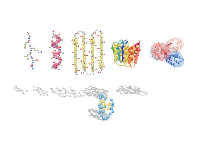
A protein starts as a long chain of building blocks called amino acids, strung together in a particular order. The chain coils into spirals and pleated sheets, which then pack into a compact 3D shape — and some proteins join with others into bigger teams. The shape decides the job: carrying oxygen, speeding up reactions or building muscle.
A protein’s amino-acid sequence (primary structure) folds into local helices and sheets (secondary), a compact three-dimensional domain (tertiary) and, for many proteins, an assembly of several chains (quaternary). The native fold is a marginally stable free-energy minimum, set by the hydrophobic effect, hydrogen bonding and the narrow range of backbone angles a planar peptide allows.
Key facts
- Peptide bond
- C–N 1.33 Å; planar, ω ≈ 180° (trans)
- α-helix
- φ ≈ −60°, ψ ≈ −45°; 3.6 res/turn; 1.5 Å rise; 5.4 Å pitch
- β-strand
- φ ≈ −120 to −140°, ψ ≈ +120 to +135°; ~3.3–3.5 Å per residue
- Helix H-bond
- C=O(i) ··· H–N(i+4), N···O ≈ 2.9–3.1 Å
- Typical ΔG of folding
- ≈ −20 to −60 kJ mol⁻¹ (marginal stability)
- Haemoglobin
- α₂β₂, 141 + 146 residues per αβ pair, ≈ 64.5 kDa
Parts you can explore (11)
- Primary structure
- Peptide planes
- α-helix
- Helix H-bonds
- β-sheet
- Sheet H-bonds
- Tertiary fold
- Hydrophobic core
- Quaternary assembly
- Haem groups
- Folding chain
In the 3D model
Rotate and take apart the model, follow a guided tour of what each part does and why it’s needed, change it with a live slider, and test yourself with a short quiz. Free, no sign-up.
Open Levels of protein structure in 3D →
OpenArc Models’ models are simplified teaching schematics. Terms · Privacy