Electromagnetism & electronics
MRI scanner
Electromagnetism · Medical imaging
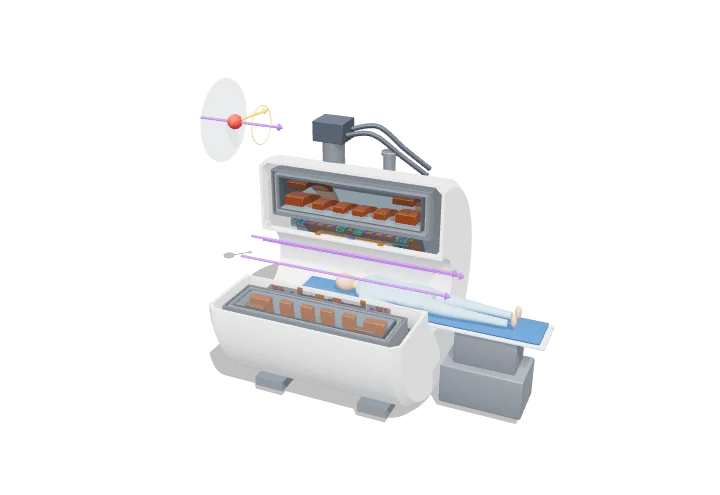 Open the interactive 3D model →
How can a scanner see inside your brain using only magnets and radio waves?
Your body is full of water, and every water molecule contains hydrogen atoms whose centres behave like tiny spinning magnets. An MRI scanner puts you inside a giant magnet that makes those tiny magnets wobble at a precise rate, then nudges them with radio waves and listens to the faint radio “echo” they send back. A computer works out where each echo came from and builds a picture.
An MRI scanner places the body in a strong, extremely uniform magnetic field from a superconducting magnet, so that hydrogen nuclei precess at the Larmor frequency f = γB₀/2π. Radio-frequency pulses tip their magnetisation, switched gradient fields encode position in the frequency and phase of the returning signal, and a computer reconstructs images from it.
Key facts
- Clinical field strengths
- 1.5 T and 3 T (7 T for research and some clinical use)
- ¹H gyromagnetic ratio γ/2π
- 42.58 MHz T⁻¹
- Larmor frequency
- 63.9 MHz at 1.5 T; 127.7 MHz at 3 T
- Coil temperature
- ≈ 4.2 K in liquid helium (NbTi T_c ≈ 9 K)
- Field uniformity
- a few ppm over a ≈ 40–50 cm sphere
- Gradient strength
- ≈ 30–80 mT m⁻¹ on clinical systems
Parts you can explore (13)
- Bore and covers
- Cryostat
- Liquid helium
- Main magnet coils
- Active shield coils
- Cold head
- Gradient coil X
- Gradient coil Y
- Gradient coil Z
- RF body coil
- Main field B0
- Patient table
- Proton spin (inset)
In the 3D model
Rotate and take apart the model, follow a guided tour of what each part does and why it’s needed, change it with a live slider, and test yourself with a short quiz. Free, no sign-up.
OpenArc Models’ models are simplified teaching schematics. Terms · Privacy