Human body
Intestinal villi
Anatomy · Digestive system
 Open the interactive 3D model →
How does a gut only about 3 cm wide fit an absorbing surface the size of a studio flat?
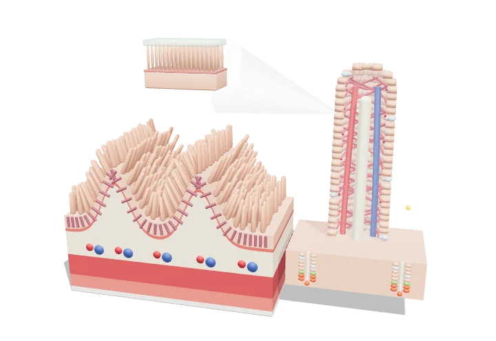
Your small intestine soaks up nutrients from digested food. Its lining is folded again and again: big folds, tiny finger-like villi on the folds, and even tinier microvilli on every cell. All that folding gives food far more surface to be absorbed through, and sugars then pass into blood vessels while fats go into lymph vessels.
The small intestine absorbs almost all digested nutrients through a wall folded at three scales — circular folds, villi and microvilli — that greatly enlarge its surface. Each villus contains capillaries that carry sugars and amino acids to the liver and a lacteal that carries absorbed fat into the lymph, while crypts at its base continuously renew the epithelium.
Key facts
- Small intestine length
- ≈ 3–5 m in vivo (≈ 6–7 m relaxed at autopsy)
- Villus height
- ≈ 0.5–1 mm (tallest in the jejunum)
- Microvillus size
- ≈ 1–2 µm long, ≈ 0.1 µm wide; ≈ 1000–3000 per cell
- Mucosal surface area
- ≈ 30 m² for the small intestine (modern morphometry)
- Epithelial turnover
- ≈ 3–5 days
- Fluid absorbed by the small intestine
- ≈ 6.5–7.5 L/day of ≈ 9 L entering
Parts you can explore (15)
- Circular folds (plicae)
- Villi
- Enterocytes
- Goblet cells
- Brush border (inset)
- Lacteal
- Capillary network
- Crypts of Lieberkühn
- Lamina propria
- Muscularis mucosae
- Submucosa
- Circular muscle layer
- Longitudinal muscle layer
- Serosa
- Nutrient absorption
In the 3D model
Rotate and take apart the model, follow a guided tour of what each part does and why it’s needed, change it with a live slider, and test yourself with a short quiz. Free, no sign-up.
OpenArc Models’ models are simplified teaching schematics. Terms · Privacy