Human body
Ear and cochlea
Anatomy · Special senses
 Open the interactive 3D model →
How does a snail-shaped tube smaller than a pea tell a bird’s chirp from a bass drum?
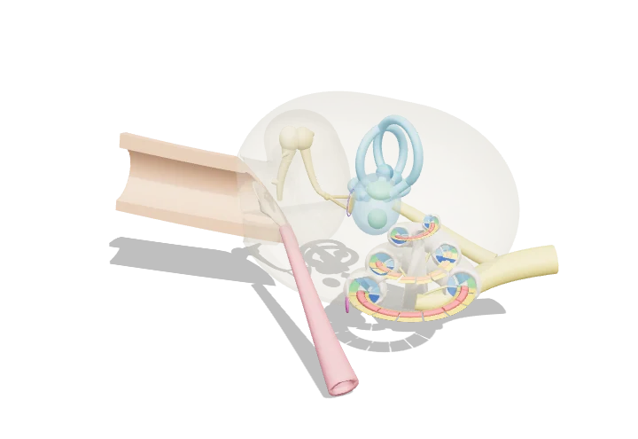
Sound waves make your eardrum shake, and three tiny bones pass that shaking into a fluid-filled spiral called the cochlea. Inside, a long membrane ripples: high sounds make it wobble most near the start, low sounds near the far end. Hair cells at each spot turn that wobble into nerve signals, so the brain knows the pitch from where the signal came from.
The outer and middle ear collect airborne sound and, through the lever and area ratio of the eardrum and ossicles, transfer it efficiently into the fluid of the cochlea. Inside the cochlea a travelling wave on the graded basilar membrane peaks at a place set by frequency, where hair cells of the organ of Corti convert motion into nerve impulses.
Key facts
- Audible range (young adult)
- ≈ 20 Hz – 20 kHz
- Basilar membrane length
- ≈ 33–35 mm; ≈ 0.1 mm wide at the base, ≈ 0.5 mm at the apex
- Cochlear turns
- ≈ 2.5–2.75
- Hair cells per cochlea
- ≈ 3,500 inner and ≈ 12,000 outer
- Middle-ear pressure gain
- ≈ 20–25× (≈ 25–30 dB)
- Endocochlear potential
- ≈ +80 mV in the scala media
Parts you can explore (16)
- Ear canal
- Tympanic membrane
- Malleus
- Incus
- Stapes
- Oval window
- Round window
- Cochlea
- Scala vestibuli
- Scala media
- Scala tympani
- Organ of Corti & basilar membrane
- Vestibule
- Semicircular canals
- Auditory nerve
- Eustachian tube
In the 3D model
Rotate and take apart the model, follow a guided tour of what each part does and why it’s needed, change it with a live slider, and test yourself with a short quiz. Free, no sign-up.
OpenArc Models’ models are simplified teaching schematics. Terms · Privacy