Cells & molecules
Haemoglobin
Biochemistry · Protein quaternary structure
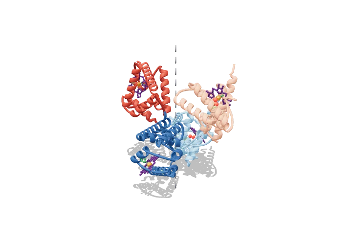 Open the interactive 3D model →
How does one protein grab oxygen tightly in your lungs and then let it go in your muscles?
Haemoglobin is the protein that makes blood red and carries oxygen around your body. It is built from four chains, each holding a flat ring with an iron atom at its centre, and each iron can hold one oxygen molecule. The clever part is teamwork: once one chain picks up oxygen, the whole protein changes shape so the other three grab theirs much more easily — and in tissues, the reverse happens.
Haemoglobin is an α2β2 tetramer whose four haem groups bind oxygen cooperatively, giving a sigmoidal saturation curve that loads O₂ in the lungs and unloads it in tissues. Cooperativity comes from a switch between a low-affinity T state and a high-affinity R state, in which one αβ dimer rotates about 15° relative to the other.
Key facts
- Composition
- 2 α chains (141 aa) + 2 β chains (146 aa) + 4 haems
- Molecular mass
- ≈ 64.5 kDa
- P50 (adult, pH 7.4, 37 °C)
- ≈ 26–27 mmHg
- Hill coefficient
- ≈ 2.7–3.0
- O₂ capacity
- 1.34 mL O₂ per g Hb (fully saturated)
- T → R rotation
- ≈ 15° of α1β1 relative to α2β2
Parts you can explore (11)
- α1 chain
- α2 chain
- β1 chain
- β2 chain
- Haem groups
- Iron (Fe²⁺)
- Proximal histidine (F8)
- Distal histidine (E7)
- Bound O₂
- 2,3-BPG
- Twofold (dyad) axis
In the 3D model
Rotate and take apart the model, follow a guided tour of what each part does and why it’s needed, change it with a live slider, and test yourself with a short quiz. Free, no sign-up.
OpenArc Models’ models are simplified teaching schematics. Terms · Privacy