Cells & molecules
Golgi apparatus
Cell biology · Organelle
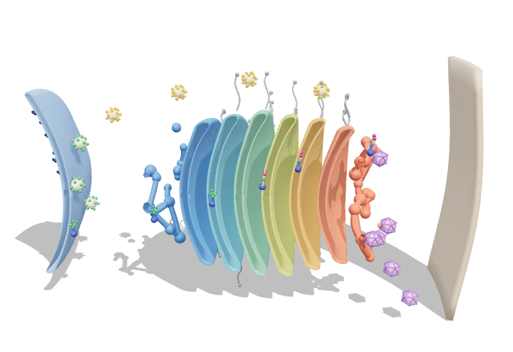 Open the interactive 3D model →
A cell makes thousands of different proteins — how does each one reach the right destination?
The Golgi apparatus is a stack of flat membrane sacs that works like a sorting office with a finishing line. Newly made proteins arrive at one face of the stack in small bubbles, are modified step by step as they pass through, and leave from the other face in bubbles addressed to where they are needed — the cell surface, storage granules, or the cell’s recycling centres.
The Golgi apparatus receives newly made proteins and lipids from the ER, modifies them — above all their sugar chains — as they pass from its cis to its trans face, and sorts them for delivery to the cell surface, secretory granules or lysosomes. Coated vesicles (COPII, COPI and clathrin) carry material into, backward through and out of the stack.
Key facts
- Cisternae per stack
- typically 4–8 (often 5–7 in mammals)
- Stack diameter
- ≈ 1 µm; cisterna lumen ≈ 20–30 nm thick
- COPI / COPII vesicles
- ≈ 60–90 nm diameter
- Clathrin-coated vesicles
- ≈ 50–100+ nm
- Transit time (ER → plasma membrane)
- ≈ 20–60 min; Golgi itself ≈ 10–20 min
- Golgi luminal pH
- ≈ 6.7 (cis) → 6.0 (TGN)
Parts you can explore (13)
- Rough ER
- COPII vesicles
- cis-Golgi network
- cis cisternae
- Medial cisternae
- trans cisternae
- trans-Golgi network
- Secretory vesicles
- Lysosomal vesicles
- COPI vesicles
- Golgins
- Glycoprotein cargo
- Plasma membrane
In the 3D model
Rotate and take apart the model, follow a guided tour of what each part does and why it’s needed, change it with a live slider, and test yourself with a short quiz. Free, no sign-up.
OpenArc Models’ models are simplified teaching schematics. Terms · Privacy