Cells & molecules
Endoplasmic reticulum
Cell biology · Organelle
 Open the interactive 3D model →
When a cell makes a protein that must leave the cell, how does it get out?
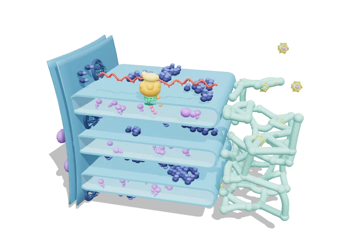
The endoplasmic reticulum (ER) is a vast folded membrane factory that fills much of a cell and joins onto the nucleus. Proteins destined for the cell surface or for export are made by ribosomes sitting on its surface and are threaded through a channel into the space inside as they are built. There they fold up, get checked, and are shipped onward in small bubbles.
The endoplasmic reticulum is a continuous network of membrane sheets and tubules, joined to the nuclear envelope, that makes most of the cell’s lipids and all of its secretory and membrane proteins. Ribosomes making these proteins dock on the rough ER and thread the growing chain through the Sec61 channel into the lumen, where it folds before leaving in COPII vesicles.
Key facts
- Share of cell membrane
- often about half of all cell membrane (≈ 50% in hepatocytes)
- Sheet lumen width
- ≈ 30–60 nm (sheets); tubules ≈ 50–100 nm diameter
- Proteins entering the ER
- ≈ one-third of human protein-coding genes
- Translation speed
- ≈ 5–6 amino acids/s in mammalian cells
- Free Ca²⁺ in lumen
- ≈ 0.1–0.8 mM vs ≈ 100 nM in cytosol
- Signal peptide length
- ≈ 15–30 residues with a 7–15-residue hydrophobic core
Parts you can explore (14)
- Nuclear envelope
- Nuclear pores
- Rough ER
- Lumen
- Bound ribosomes
- Smooth ER
- ER exit sites
- COPII vesicles
- Chaperones (BiP)
- Sec61 translocon
- Translating ribosome
- mRNA
- Signal peptide
- Nascent polypeptide
In the 3D model
Rotate and take apart the model, follow a guided tour of what each part does and why it’s needed, change it with a live slider, and test yourself with a short quiz. Free, no sign-up.
Open Endoplasmic reticulum in 3D →
OpenArc Models’ models are simplified teaching schematics. Terms · Privacy