Physics
Compound light microscope
Optics · Instruments
 Open the interactive 3D model →
How do two small lenses make a single blood cell look as big as a coin?
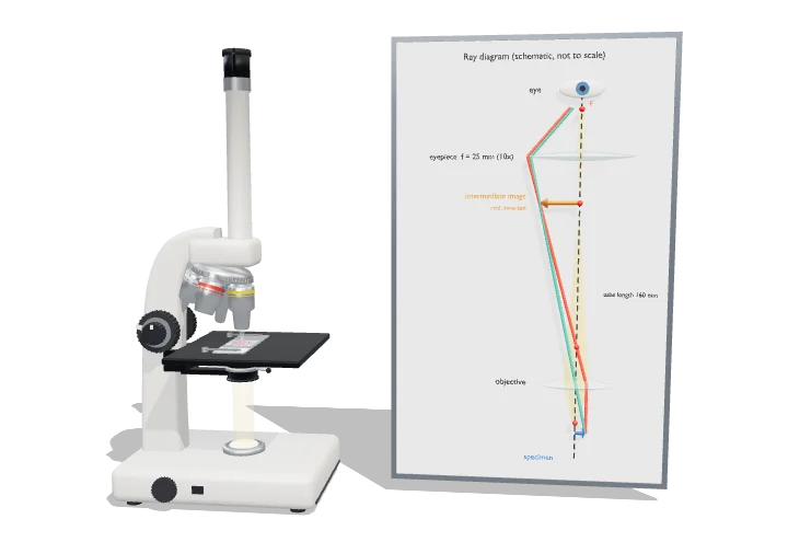
A microscope uses two sets of lenses one after the other. The lens near the slide makes a bigger picture inside the tube, and the lens you look through makes that picture bigger again. But there is a limit: past a certain point, making things bigger just makes them blurrier.
A compound microscope magnifies in two stages: the objective forms a real, inverted, magnified image inside the tube, and the eyepiece acts as a magnifier for that image. How much detail it can show is set not by magnification but by the numerical aperture of the objective and the wavelength of light, through Abbe’s limit d = λ/2NA.
Key facts
- Total magnification
- objective × eyepiece: 40× to 1000× with 4×–100× objectives and a 10× eyepiece
- Mechanical tube length
- 160 mm (classic finite-conjugate standard); modern stands use infinity-corrected optics
- Parfocal distance
- 45 mm, shoulder to specimen, so objectives stay nearly in focus when swapped
- Best resolution (visible light)
- ≈ 0.2 µm with a 1.4 NA oil objective at 550 nm
- Numerical aperture
- 0.10 (4×) to 1.25–1.4 (oil 100×); NA = n sin α
- Working distance
- ≈ 17 mm (4×) down to ≈ 0.15 mm (100× oil)
Parts you can explore (15)
- Base
- Illuminator
- Arm
- Body tube
- Eyepiece
- Revolving nosepiece
- Objectives
- Stage
- Slide
- Condenser & iris
- Focus knobs
- Light path
- Ray diagram
- Image-forming rays
- Aperture cone
In the 3D model
Rotate and take apart the model, follow a guided tour of what each part does and why it’s needed, change it with a live slider, and test yourself with a short quiz. Free, no sign-up.
Open Compound light microscope in 3D →
OpenArc Models’ models are simplified teaching schematics. Terms · Privacy