Cells & molecules
Epithelial cell junctions
Cell biology · Tissues
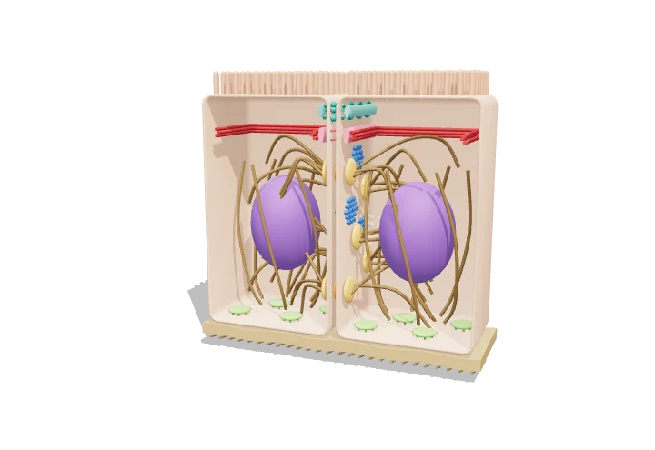 Open the interactive 3D model →
Why doesn’t your gut leak, and why doesn’t your skin peel apart when you rub it?
The cells that line your gut and cover your skin are joined to each other by tiny connectors called junctions. Some seal the gaps between cells, some act like glue or rivets, some are little tunnels between neighbours, and some anchor the cells to the layer beneath. Together they turn separate cells into a strong, leak-proof sheet.
Epithelial cells are joined by specialised junctions that seal the sheet, hold cells together, spread mechanical load, connect neighbouring cytoplasms and anchor the sheet to the basal lamina. Each junction pairs a transmembrane adhesion protein with a specific cytoskeletal partner: claudins with the ZO scaffold, cadherins with actin, desmosomal cadherins and α6β4 integrin with keratin.
Key facts
- Tight junction cleft
- ≈ 0 nm at strand contacts (outer leaflets touch)
- Adherens junction cleft
- ≈ 20 nm (15–25 nm)
- Desmosome cleft
- ≈ 30 nm, with a dense midline
- Gap junction gap
- ≈ 2–4 nm; pore ≈ 1.5 nm
- Gap junction size limit
- molecules up to ≈ 1 kDa
- Human gene families
- ≈ 27 claudins, 21 connexins, > 100 cadherin superfamily members
Parts you can explore (10)
- Epithelial cells
- Microvilli
- Tight junction
- Adherens junction
- Actin belt
- Desmosomes
- Keratin filaments
- Gap junctions
- Hemidesmosomes
- Basal lamina
In the 3D model
Rotate and take apart the model, follow a guided tour of what each part does and why it’s needed, change it with a live slider, and test yourself with a short quiz. Free, no sign-up.
Open Epithelial cell junctions in 3D →
OpenArc Models’ models are simplified teaching schematics. Terms · Privacy