Cells & molecules
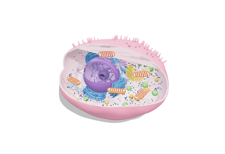
Animal cell (cutaway)
Cell biology · Eukaryotic cell
 Open the interactive 3D model →
Your body is built from about 30 trillion cells — what is going on inside each one?
An animal cell is a tiny bag of watery gel, too small to see without a microscope. Inside are compartments called organelles, each with a job: one stores the instructions, others make proteins, pack them, deliver them or supply energy. Together they work like a busy factory.
An animal cell is a membrane-bounded compartment, typically 10–30 µm across, subdivided into organelles that separate incompatible chemistries such as DNA replication, protein secretion, ATP synthesis and digestion. Proteins made on ribosomes are routed through the ER, Golgi and vesicles, while a cytoskeleton of microtubules, actin and intermediate filaments gives shape and serves as tracks for motor-driven transport.
Key facts
- Typical diameter
- ≈ 10–30 µm (volume ≈ 1–4 pL)
- Plasma membrane thickness
- ≈ 5 nm
- Nucleus diameter
- ≈ 5–10 µm; ≈ 3,000–4,000 nuclear pores
- Mitochondria per cell
- ≈ hundreds to thousands (≈ 22% of a hepatocyte’s volume)
- Protein concentration
- ≈ 200–300 mg/mL
- Lysosome lumen pH
- ≈ 4.5–5.0 (cytosol ≈ 7.2)
Parts you can explore (13)
- Cell membrane
- Cytosol
- Nucleus
- Nucleolus
- Mitochondria
- Endoplasmic reticulum
- Golgi apparatus
- Ribosomes
- Vesicles
- Lysosomes
- Peroxisomes
- Centrosome
- Cytoskeleton
In the 3D model
Rotate and take apart the model, follow a guided tour of what each part does and why it’s needed, change it with a live slider, and test yourself with a short quiz. Free, no sign-up.
Open Animal cell (cutaway) in 3D →
OpenArc Models’ models are simplified teaching schematics. Terms · Privacy